Category Archives: Growth

Cell Walls

One of the most prominent features of plant cells is their cell wall. While many organisms produce some kind of extracellular matrix that is important in cell function, almost every cell made by a plant is surrounded by a rigid mixture of cellulose and other polysaccharides and proteins. The cell wall allows the formation of high hydrostatic pressure known as turgor pressure.

Cellulose

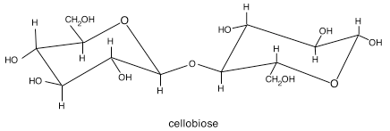
Cellulose is the major load-bearing structural component of the cell wall. In chemical terms, cellulose is β1→4-linked glucose dimers called cellobiose. These dimers repeat in long chains, which are crystallized with other such chains during synthesis to form microfibrils. Think of a microfibril as yarn, and individual chains as the threads that are spun into a yarn.

chemical structure of cellobiose

One of the peculiarities of cellulose in the cell wall is that it is not synthesized inside the cell and deposited in the apoplast by vesicles. Rather, cellulose is synthesized by a complex spanning the plasma membrane and extruded into the apoplast. These rosette-shaped structures, called terminal complexes, are made up of many individual cellulose synthase enzymes working in concert to produce a microfibril. Terminal complexes travel along a network of cortical microtubules that determine the pattern of deposition of cellulose microfibrils. Direct evidence for this hypothesis came from visualizing the interaction between microtubules and a part of the cellulose synthase enzyme complex.

microfibril-synthesis

Other Wall Components

In addition to cellulose, plant cell walls include a wide variety of polysaccharides and structural proteins. The polysaccharides include hemicelluloses and pectins, both of which are diverse groups of complex, branched carbohydrates. Unlike cellulose, neither of these constituents is manufactured directly in the cell wall, but both follow the more traditional intracellular synthesis followed by secretion. These polysaccharides function to stabilize the interaction between cellulose microfibrils through H-bonding.

cartoon diagram of cell wall components

Pardez AR, Somerville CR, Ehrhardt DW (2006) Visualization of Cellulose Synthase Demonstrates Functional Association with Microtubules. Science 312:1491-1495 See a collection of time-lapse movies.

Growth

How do plants grow?

  • Principles of Growth: How do plants create a complex body plan?
  • Meristems: How do these specialized regions of cell division make a plant?
  • Cell fate determination: Do cells inherit their identity, or is it the product of position?
  • Cell walls: What role do cell walls play in growth and development?
  • Cell expansion: What controls the timing and direction of cell expansion?
  • Water potential: How does water provide the driving force for cell growth?
  • Organ formation: How does a jumble of cells become organized for a specific function?

Organ Formation

Although the most primitive land plants possessed little more than stem-like structures, most land plants today produce broad, flat leaves that maximize the surface area exposed to light. While it may be clear how meristem-driven growth can result in the formation of an axial organ, like a stem or root, it is probably harder to imagine how a meristem could produce a laminar structure like a leaf.

The first visible indication that a leaf is about to form is a tiny bump on the surface of the apical dome, the tip-most section of the elongating stem. These bumps form off to one side of the dome, and the position of the bump determines the pattern of leaf placement along the stem, called phyllotaxy. In other words, for a species having alternate phyllotaxy, the position of consecutive bumps would spiral around the apical dome, while species with opposite phyllotaxy would form two bumps simultaneously and offset by 180° from each other. As time passes, the bumps, known as leaf primordia, expand into leaves, and the leaves become separated by expansion of the stem over the time between which the bumps formed.

Given that the arrangement of leaves on a stem is highly reproducible within a species, the placement of each leaf primordium must be regulated. Over the last 20 years, there has been substantial progress made in identifying some of the factors that control maintenance of the meristematic population as well as initiation of new leaf primordia. One of the key concepts to emerge is that, while the meristem looks like an amorphous mass of cells, it is actually organized into two zones based on the expression patterns of particular classes of genes, many of which encode transcription factors. These two zones, known as the central zone (CZ) and peripheral zone (PZ), exist in a dynamic tension, such that they tend to protect their cellular ‘turf’ by preventing expression of genes from the other zone. In addition to zones, the shoot apical meristem is also partitioned into layers, with most species having 3 layers (L1, L2, and L3).

Leaf primordia form in the peripheral zone, and even before a bump is visible, the region where a primordium will form shows downregulation of a transcription factor associated with meristem activity, the STM gene in Arabidopsis. Shortly thereafter, this region begins to protrude from the surface as a leaf primordium. The L1 layer, which is the outermost layer of cells, will form the dermal tissue of the new leaf, while the L3 layer will form the vascular tissues. L2 goes on to differentiate into the rest of the tissues that make up the leaf, most of which are ground tissue. As the leaf primordium is beginning to emerge, cell divisions in L1 and L2 occur perpendicular to the surface of the apex, while those in L3 occur in both perpendicular and parallel planes.

In addition to an important role for transcription factors and signal peptides, plant hormones also have prominent roles in the emergence of leaf primordia. Using an auxin-responsive reporter gene, it has been determined that leaf primordia are local sites of auxin maxima. Auxin, or indole-acetic acid (IAA) is synthesized in existing primordia and transported across the apical dome, where it appears to accumulate and coincide with the site of primordium formation. This accumulation requires a system of polar auxin transporters, which have now been shown to direct auxin transport across the shoot apex to the site of incipient primordium formation. As the new primordium emerges, it exports the auxin necessary to form a new maximum on the other side of the meristem, where a new auxin maximum will form. In other words, as primordia mature, they become auxin sources rather than auxin sinks.

Reference
Barton MK (2010) Twenty years on: The inner workings of the shoot apical meristem, a developmental dynamo. Developmental Biology 341:95-113

Water Potential

For cell expansion to continue, cells must continuously take up water from their surroundings. The movement of water occurs in response to several forces, including its concentration, pressure, position in the gravity field, degree of association with soil particles, etc.

Kinds of water flow

The driving force for much of water’s movement at the cellular level is diffusion, the random intermingling of molecules as they collide with like molecules and change directions. Because of these collisions, over time, diffusion tends to result in the movement of molecules from an area of higher concentration to an area of lower concentration. The force behind such a directional movement is therefore the concentration gradient of the molecule. While we are accustomed to thinking about the concentration of a solute, such as a salt, dissolved in a solvent, it is also just as appropriate to speak in terms of the concentration of the solvent itself, as we will see.

Diffusion is a very effective means of movement, but only across short distances or within small volumes. For example, a small molecule in aqueous solution can diffuse 50 μm in about 0.6 s. However, for that same molecule to diffuse 1 m would require 2.5 x 10⁸ seconds, or about 8 yrs! Thankfully, plants are not limited to diffusion as a means of water transport, but also employ bulk flow, which is the movement of a mass of molecules, often in response to a pressure gradient. This kind of transport is employed by the vascular tissues of the plant to accomplish transport across great distances.

A special kind of diffusion called osmosis occurs in living systems, when a selectively permeable membrane is involved. Very often, forces besides solvent concentration influence osmotic flow, with the sum of all such forces represented by the free energy of the solvent. If diffusion is in response to a concentration gradient, and bulk flow a pressure gradient, osmosis is in response to the sum of both of these forces and other influences, all of which together describe water potential.

Water Potential

Water potential is a term denoting the chemical potential of water, which is to say the free energy of water — the energy available to do work. This energy is expressed in units of energy per unit volume J m-3, which is equivalent to pressure (in MPa). There are many contributing factors to water potential, as we’ve seen above. Always keep in mind that pure water has a water potential of 0 (\Psi_w = 0)

The solute potential (\Psi_s) represents the effect of any solutes on the free energy of water. The addition of a solute to water decreases the concentration of water, thus reducing its free energy. The degree of this reduction is a function of the final concentration of solute particles:

\Psi _s = -cRT

where c is in units of osmolality (moles of total dissolved solutes per L). Note the negative sign, a reminder that solutes lower water potential.

The pressure potential (\Psi_p) represents the effect of pressure on the free energy of water; positive pressure increases free energy, and negative pressure reduces it. Pressure within a typical leaf or root cell is positive, while that within the xylem is negative. A cell with \Psi_p = 0 is flaccid.

For some tall plants, gravity becomes an important consideration in predicting total water movement, thus it is necessary to consider the gravity potential (\Psi_g) in such a plant. Each 10 m in vertical distance translates to a 0.1 MPa change in water potential.

Under some conditions, the matric potential (\Psi_m) plays a significant role in determining the flow of water. When water is bound to a surface in a very thin layer, that water is less available to do work, reducing its free energy. This parameter plays an important role during seed germination and under drought conditions.

The effect of all of these factors on the overall water potential is additive, and can be expressed as follows:

\Delta \Psi_w = \Delta \Psi_s + \Delta \Psi_p + \Delta \Psi_g + \Delta \Psi_m

Under most circumstances, the first two terms, corresponding to solute and pressure potentials, are sufficient to determine plant water potential. Plant water potential governs transport of water across membranes, with water always moving from an area of high to low water potential.

Cell expansion

It should be clear by now that the addition of new cells by division in meristematic zones represents an important component of growth. But the dividing of a mother cell in two does not necessarily result in an increase in size. For such an increase to occur, division must be coupled with cell expansion. Without expansion, a dividing cell is just partitioned into smaller and smaller elements.

The force that powers cell expansion is turgor pressure, a positive hydrostatic pressure that builds up within the cell due to the uptake of water. A typical leaf cell in a well-watered plant has a turgor pressure between 0.1 and 3.0 MPa (MPa = megapascals). For reference, a typical car tire is inflated to 30 psi (pounds per square inch), which is equivalent to 0.2 MPa. While such a tremendous pressure would cause most cells to burst, plant cells maintain such high pressures through the resistance of the cell wall.

Cell expansion, then, represents a carefully-controlled balance between wall resistance and wall yielding. For each cell, there is a minimum of pressure required to allow expansion, and above that minimum, expansion proceeds depending on the wall extensibility:

\frac{dV}{dt} = m (P - Y)

where the rate of change in volume, \frac{dV}{dt}, is equal to the product of effective pressure (turgor pressure (P) minus wall yield threshold (Y)) and wall extensibility (m). This is a version of the Lockhart equation, first published in 19651.

Of these factors, the cell has direct temporal control over wall extensibility. One means of regulation over extensibility is by acidification of the cell wall through proton pumping. Lowering the pH of the wall is associated with increased extension. Another means of control is through the production, secretion, and stimulation of wall-bound proteins that loosen bonds between the structural components of the wall. Foremost among these is a family of proteins called expansins, that appear to temporarily loosen the non-covalent linkages between cellulose and other wall polysaccharides. By loosening these linkages, expansins allow the load-bearing members of the wall to slide past each other and permit cell expansion in a very controlled manner.

The wall loosening carried out by the expansins takes place in specific wall locations under specific conditions, allowing the cell to control the timing of expansion and ultimate shape of the cell. Without such control, cells would be free to expand in any direction and at any time, which would have clear implications for tissue and organ form in the mature plant. In addition, the direction of cell expansion is heavily influenced by the pattern of deposition of cellulose microfibrils, which can also serve to constrain the kinds of final shapes a particular cell can achieve.

There is a significant brake in this system of turgor-driven cell expansion, and that is a constraint imposed by biophysics. As turgor pressure drives cell expansion, the volume of the cell increases. This increase in volume reduces the turgor pressure pushing on the wall and providing the force for expansion. Because even a slight increase in volume reduces turgor pressure, water must be taken up continuously to maintain the turgor pressure required to sustain growth.


  1. Lockhart, J.A. (1965) An analysis of irreversible plant cell elongation. Journal of Theoretical Biology 8:264-275. 

Cell fate determination

The question of what the derivative cell will become after it leaves the meristem is governed by its position within the organ. The question of cell fate has been of interest to plant scientists for centuries, but there was little way to test the competing hypotheses of cell lineage and cell position. In an elegant series of experiments, researchers have shown that plant cells can change their fate by being forced into a new position, taking their cues for which kind of cell to form based on their neighbors rather than the identity of their mother cell. Since that set of experiments, researchers have started to identify the signals that transmit this identity information between adjacent cells, largely through microscopic analysis of mutants.

At the broad end of the spectrum, all plant cells fall into one of three tissues with respect to identity: vascular, dermal, and ground tissues. Vascular tissue is composed of cells involved in transport and cells that support transport. Dermal tissue is composed of cells on the surface of the plant. Ground tissue is composed of cells that make up the rest of the plant – in the interior of leaves, stems, and roots but not having a specific role in transport. Thus, the fate of any particular derivative to become a cell in either of these three tissue systems is determined by the location the derivative finds itself within the meristem.

Because of the centrality of position in determining cell fate, one of the most important facets of plant development is the specification of the plane of cell division. For any given mother cell undergoing division, the orientation of the plane of division could mean the difference between a derivative becoming an epidermal cell (dermal tissue) or a cortex cell (ground tissue). This is the case not only because plant cells don’t migrate and adhere, and not only because of the primary role of position in determining cell fate, but also because of the presence of the thick, rigid, cellulosic cell wall. Once wall deposition begins during cytokinesis, there is no chance for a cell to change its position, and hence its fate.

Within the meristem, cells pass through the cell cycle with relatively high frequency. While the process of mitosis has some notable differences from other eukaryotic cells, cytokinesis in plant cells is almost wholly unlike that seen in other eukaryotes. The “textbook” cell undergoes cytokinesis through the familiar ‘pinching in’ of the plasma membrane, but the rigid wall of plant cells is not conducive to such a process. Rather, cytokinesis progresses by the deposition of polysaccharides and membrane lipids at the site of the new cell wall through vesicle trafficking. These vesicles travel along a specialized arrangement of microtubules known as the phragmoplast. Imagine dividing a room in two by suspending a piece of plaster in mid-air, then adding to it until you had built a complete wall, and you would have some idea of how cytokinesis in plants occurs.

Meristems

Although the plant emerges from embryogenesis with few outwardly visible structures, there is nonetheless already a significant amount of organization in place in the plant embryo. Chief among the organizing structures are two apical meristems, one at the shoot tip and another at the root tip. From these two regions will come all future branches, roots, leaves, and flowers.

light micrograph of the outline of root cells
Root tip of Arabidopsis thaliana, showing the organization of cell files in distinct lineages from the meristem population.

Apical meristems are collections of dividing, undifferentiated cells. When a meristematic cell divides into two daughter cells, one of them is known as an initial, and the other is known as a derivative. Assuming the meristem is neither increasing or decreasing in size over time, only one of these two daughter cells – the initial – will remain in the meristem. The derivative cell will “leave” the meristem eventually, not by cell migration as seen in animals, but by being pushed farther and farther from the meristem proper by continued rounds of division and expansion. As you study the image of a root apex at right, notice how cells form files that almost seem to ‘flow’ from the tip toward the base.

Meristems are therefore a source of new, undifferentiated cells in the tips of growing stems and roots. As they are pushed out of the meristem through division and expansion, these ‘blank slate’ cells begin to take on a cell identity as one of the three major tissue types: dermal, ground, or vascular. In addition to adopting a cell identity, each cell takes on a shape appropriate for its type and location. The shape of individual cells provides the driving force for the formation of organs like leaves or petals.

Principles of Growth

Plants follow a fundamentally different process for growth than animals. For example, it would be odd (to say the least) to find a new limb forming on a person after embryonic development, yet plants produce nearly all of their form post-embryonically, with only a minimal set of body parts created during embryogenesis. The ability to continue producing organs after embryo formation demonstrates that much of plant growth is indeterminate, meaning a plant can continue producing new organs like leaves over and over. This strategy has proven to be flexible and adaptable across a wide range of ecosystems, allowing some plants to live for hundreds, or even thousands, of years.

Ginkgo embryo and gametophyte

In addition to indeterminate growth, plants also show a high degree of developmental plasticity. For an organism that is not free to run away from drought, cold, or herbivores, this attribute is an important strategy. No two plants ever look exactly the same because of this plasticity, and this property allows individuals to become tailor-made for their specific site, producing roots in just the right locations, avoiding rocks and other roots, and sending out branches with leaves right where they will intercept light.

There are, however, certain patterns that seem to be repeated when one looks closely at a plant. The spacing of leaves along a stem, for example, tends to follow a fairly repeatable pattern. This and other patterns indicate that development in plants is iterative, suggesting a pre-recorded program that repeats itself after a certain period.

Plant growth follows these three principles – it is indeterminate, plastic, and iterative. The application of these principles leads to the tremendous variety of forms we find in the plant kingdom, from the arrangement of petals in a flower to the spiral alignment of branches on a pine tree. And all of these arise from the organization of cells into form-producing factories known as meristems.