The carbon reactions of photosynthesis, also known as the Calvin-Benson cycle or photosynthetic carbon reduction, consist of a series of steps that take place in the chloroplast. Broadly speaking, the carbon reactions consist of three steps: carbon fixation, reduction, and regeneration of the initial substrate of the cycle. Because the carbon reactions together form a metabolic cycle, there is no end product of the pathway as such. Rather, the cycle ‘exports’ reduced carbon in the form of triose phosphates.
Carboxylation
In the first step of the carbon reactions, CO2 reacts with a high-energy five carbon substrate called ribulose bisphosphate (RuBP), resulting the the formation of two molecules of the three carbon compound phosphoglycerate (PGA). This carboxylation reaction is catalyzed by Rubisco, a large and evolutionarily ancient enzyme made up of 8 large and 8 small subunits. Rubisco is one of the most abundant proteins on Earth, and makes up 40% of the soluble protein in an average leaf. Rather than attaching the CO2 onto the end of the five carbon substrate, Rubisco adds it to the second carbon in the chain, forming a very unstable intermediate that splits between the 2 and 3 carbons almost immediately. The resulting PGA molecules enter the reduction phase of the cycle.
Reduction
The first step of the reduction phase of the carbon reactions is where the majority of the energetic compounds from the light reactions are used. In the first reaction of reduction, PGA is phosphorylated to make bisphosphoglycerate, a reaction that uses ATP produced by the light reactions. In the second reaction of the reduction phase, bis-phosphoglycerate is reduced to glyceraldehyde-3-phosphate (G3P) by the donation of electrons from NADPH, also a product of the light reactions. G3P is a triose phosphate carbohydrate and is the true product of the carbon reactions in the sense that it is exported from the cycle and enters the starch or sucrose biosynthetic pathways.
Regeneration
The rest of the carbon reactions, 10 individual enzyme-mediated reactions, make up the regeneration phase of the cycle. The complexity of the regeneration phase is due to the difficult task it is given: to make a highly reactive five-carbon molecule starting with a three-carbon molecule. To accomplish this, the regeneration phase employs a network of interrelated reactions in which the pool of triose phosphates serve as reactants many different times.
Regulation
The carbon reactions are regulated by two key factors: the pH of the stroma and a redox regulatory feedback loop. Although the carbon reactions are sometimes referred to as the ‘dark reactions’ to illustrate their separation from the light reactions, this is a misnomer. The carbon reactions are highly dependent on light, not just for the products of the light reactions that act as substrates, but also for regulation of several key enzymes. Three enzymes in particular have a pH optimum around 8, including Rubisco (the other two are fructose 1,6-bisphosphatase and sedoheptulose bisphosphatase). This high pH requires the transport of protons out of the stroma and into the lumen, thus the maximum activity of these enzymes occurs in the light.
A second point of regulation of the carbon reactions is a redox regulatory feedback loop. If you recall, PS I reduces an iron-sulfur protein called ferredoxin (Fd), which can supply electrons to NADP+. In addition, it can reduce another small redox-active protein called thioredoxin. When reduced, it can reduce disulfide bridges that exist in several enzymes in the carbon reactions, changing them from an inactive state to an active state.
Efficiency
To calculate the overall efficiency of photosynthesis, we need to correlate the input energy from light with the output energy in the form of carbohydrates. On average, it takes 8 photons of light to fix one molecule of CO2 via the reduction of 2 NADP+ molecules and the synthesis of 3 ATPs. Thus it will require an input of 48 photons to fix one molecule of a hexose carbohydrate (six CO2 fixation events). If we assume each of these photons contains the minimum energy necessary to drive photosynthesis, about 680 nm light, that would be 175 kJ per mol of photons. Therefore, multiplying 48 mol × 175 kJ per mol gives 8400 kJ of energy for 1 mole of hexose carbohydrate. This represents the minimum energy required by the plant to produce a mole of hexose. When fully oxidized by the cell, this carbohydrate yields 2804 kJ of energy, giving an overall theoretical efficiency of 33% (2804 / 8400).
The photosynthetic light reactions take place across the thylakoid membrane, an extensive system of membranes extending throughout the chloroplast. This network of membranes is home to a variety of membrane-spanning proteins involved in the interception of light or the transfer of electrons. Working together, these proteins transduce the energy of light into an electrochemical gradient across the thylakoid that ultimately drives ATP synthesis. At the same time, they form a series circuit that removes electrons from water and donates them to an electron acceptor called NADP+, producing NADPH.
Light Harvesting
While there is a variety of pigment molecules involved in photosynthesis, light harvesting and energy transduction are carried out by the chlorophylls. Chlorophylls are closed-ring tetrapyrroles that coordinate a Mg+ in the center of the ring that plays a role in the redox reactions at the core of photosynthesis. In addition, they have a long fatty-acid tail that renders them hydrophobic. Chlorophylls interact with light and show two peaks in their absorption spectra: one in the red range and another in the blue.
Chlorophylls are not freely distributed in the thylakoid membrane, but are found in association with polypeptides. One such class of protein is known as the Light Harvesting Complex, or LHC proteins. These proteins act as antenna complexes, gathering light energy and directing it to the reaction center chlorophylls. In other words, the chlorophylls associated with the antenna complex proteins are not themselves chemically active — that role is reserved for the reaction center chlorophylls. On average, only 1 in 250 chlorophyll molecules is chemically active.
The chlorophylls associated with antenna complex proteins are nonetheless critical to photosynthesis, because as they are struck by photons and excited, they are capable of transmitting that excitation to nearby chlorophylls through resonance energy transfer. Research has shown that this process of energy transfer is an example of quantum coherence, and the network of chlorophylls acts in concert to transfer excitation energy with near 100% efficiency to the reaction center chlorophylls.
Reaction Centers
The LHC proteins surround photosynthetic reaction center proteins. The reaction center proteins also have a number of chlorophylls associated with them that receive the excitation energy from LHC chlorophylls. In addition, each of the 2 reaction center complexes has a set of chlorophylls that are chemically active — they are capable of photochemistry. These chlorophylls, known as the reaction center chlorophylls or special pair, transfer an electron to an electron acceptor upon excitation. When in the ground state, prior to absorbing light energy, the reaction center chlorophyll special pair is both a poor reducing agent and a poor oxidizer. But when excited by energy absorbance, it becomes both a good reducing agent and oxidizer.
Evidence for two photosynthetic reaction centers in plants came from a series of experiments carried out by Robert Emerson. Working with the green alga Chlorella, Emerson used flashes of light at specific wavelengths to drive photosynthesis while measuring the net yield of O2. He observed a particular rate of O2 yield under both 670 nm and 700 nm light. When the culture of algae was excited by both wavelengths simultaneously, however, Emerson measured a rate of O2 yield that was greater than the sum of O2 produced by either wavelength alone. This simple experiment had profound implications, as Emerson and many others would go on to show that photosynthetic electron transport occurred through a series of two reaction centers in series. In other words, electrons passed from one of the reaction centers to the other through a number of intermediaries in and around the thylakoid. The two photosynthetic reaction centers are known as Photosystem II (PS II) and Photosystem I (PS I).
Photochemistry
The excitation of the reaction center chlorophyll special pair in PS II raises the energy level of this molecular complex to the point that it becomes energetically favorable for it to lose an electron. As a result, the excited-state chlorophyll reduces a nearby molecule of pheophytin, which is nearly identical in structure to chlorophyll save for the Mg+ at its center. This structural similarity ‘tunes’ the pheophytin to be an efficient electron acceptor from the excited state special pair. The positioning of the primary acceptor pheophytin is important as well, as it is found in the reaction center protein toward the stromal face of the membrane spanning domain. This position results in the charge separation across the thylakoid membrane that underlies the production of an electrochemical gradient.
Midpoint potentials of the various redox components in photosynthetic electron transport.
The loss of an electron from the reaction center special pair is known as primary photochemistry, and this leaves the special pair in an unstable, oxidized state. In PS II, the special pair can be re-reduced by a nearby tyrosine amino acid, leaving the tyrosine oxidized. If we follow the trail of redox reactions back to their origin, we find that ultimately, the electrons that re-reduce the special pair are removed from water at a polypeptide closely associated with the lumenal face of the PSII reaction center called the Oxygen Evolving Complex (OEC).
The OEC catalyzes the oxidation of water through a stepwise series of redox intermediate states made possible through its coordination of Mn2+ ions. The OEC thus provides electrons one-at-a-time to the reaction center special pair as they are removed from a cluster of Mn2+ ions. These electrons are eventually replaced on Mn2+ when two water molecules are simultaneously oxidized, releasing four protons (H+) and molecular oxygen (O2). In other words, the oxidizing potential of the reaction center special pair is transferred to the OEC, where it is used to oxidize water.
In PS I, the mechanics of photochemistry occur the same as on PS II, but the primary electron acceptor is a modified chlorophyll rather than pheophytin. Rather than having an OEC as a source of electrons, the electron donor for PS I is a small molecule called plastocyanin, which diffuses to the lumenal face of PS I carrying electrons that ultimately came from water via PS II.
Electron Transport
After the oxidation of the PS II reaction center, the lost electron enters a pathway of electron flow through several complexes embedded in the thylakoid membrane. This electron transport pathway includes the protein complexes PS II, cytochrome b6f, and PS I. While it is convenient to represent each of these three components nearby each other, they are actually localized in different parts of the thylakoid membrane. PS II and cytochrome b6f are in the interior portions of thylakoid stack, while PS I is found in regions of the membrane having access to the stroma. Two pools of electron carriers connect each of the three major components, with a small, hydrophobic organic molecule called plastoquinone diffusing between PS II and cytochrome b6f, while a copper-containing protein called plastocyanin links cytochrome b6f with PS I. In other words, PS II reduces plastoquinone, and plastocyanin serves as the electron donor for PS I.
The three major components of the electron transport system do no exist to simply pass electrons along to NADP. Their orientation within the thylakoid is such that, as electrons travel through the various components, their flow enhances the concentration of protons that forms in the lumen due to water oxidation. As described above, the direction of electron movement during photochemistry on PS II is from the lumenal face toward the stromal face of the thylakoid due to the position of the primary electron acceptor. This directional flow allows plastoquinone to gather two protons from the stroma along with the two electrons it receives from PS II. When reduced, plastoquinone leaves PS II and diffuses through the membrane and binds to a cytochrome b6f complex on the lumenal face. As plastoquinone reduces the cytochrome b6f complex, the two protons it was carrying are released into the lumen.
Cytochrome b6f engages in two different processes with the electrons it receives from plastoquinone: non-cyclic transport and cyclic electron transport. When performing non-cyclic transport, the cytochrome b6f complex is reduced by plastoquinone and reduces plastocyanin at the lumenal face. Under certain conditions though, cytochrome b6f passes electrons among several intrinsic electron acceptor sites, each of which can coordinate a quinone molecule. Because this process relies on quinone sites, this has been named the Q cycle. The various quinone binding sites on the cytochrome b6f complex facilitate the flow of electrons toward the stromal face, where they eventually reduce a plastoquinone associated with the cytochrome b6f complex. This plastoquinone is identical to that described above, and once reduced it leaves its binding site at the stromal face of the cytochrome b6f complex and binds at the complex’s lumenal face. The end result of the Q cycle is to further enhance the flow of protons into the lumen.
Once reduced by cytochrome b6f, plastocyanin (a single electron carrier) leaves the complex and diffuses through the lumen to PS I. As the electron donor to PS I, plastocyanin is oxidized by the PS I reaction center special pair when it absorbs a photon of light energy and undergoes photochemistry. The electron that is lost from the reaction center travels to a modified chlorophyll and several intermediate electron acceptors within PS I before reducing ferredoxin, bound to the stromal side of PS I. Ferredoxin is a small protein that coordinates an iron-sulfur complex. When reduced, it provides the reducing power to the enzyme involved in reducing NADP+, called ferredoxin-NADP reductase. It can also participate in cyclic flow of electrons between PS I and the cytochrome b6f complex under certain conditions. The net result of the electron transport pathway is the conservation of light energy in the form of an electrochemical potential gradient across the thylakoid membrane, and the reduction of NADP+ to NADPH.
ATP Synthesis
The energy that is represented by the proton gradient across the thylakoid membrane can be used by ATP synthase to catalyze the addition of a phosphate onto ADP to make ATP. The ATP synthase enzyme is a multimeric molecular machine that converts the gradient of protons into kinetic, rotational energy by permitting protons to flow through its membrane-spanning pore. As protons leave the region of high concentration in the lumen and pass through the pore region, they exit into the stroma, which causes rotation of the membrane-spanning domain. This rotation is propagated by a shaft-like domain called the γ domain to the catalytic domain. Within the catalytic domain, ADP and P(i) are bound, and the rotation of the γ domain causes a conformational change that forces the ADP and P(i) to react and produce ATP.
What we call ‘photosynthesis’ is a collection of events that uses the energy contained in light and reducing CO2. A simplified net reaction is shown below:
H2O + CO2 + light → (CH2O)n + O2
On an annual basis, this collection of processes is responsible for fixing 7 × 10¹³ kg of CO2. To help put this value in perspective, that is equal to 1% of all known fossil fuel reserves on the planet and roughly 10× the annual global energy consumption. In addition, photosynthesis also releases oxygen at a rate that replaces all O2 in the atmosphere every 2000 years.
Photosynthesis consists of 2 interdependent processes known as the light reactions and the carbon reactions. For our purposes, we will further disassemble the light reactions into the processes of photochemistry and electron transport. Both of these parts of the light reactions take place across the system of membranes found within chloroplasts, known as the thylakoid membrane system.
As we will discuss in more detail elsewhere, light results in the polarization of this membrane due to the formation of a H+ gradient. This proton gradient is used to by ATP synthase to combine ADP and Pi to form ATP, which is one of the key products of the light reactions. The other major product of the light reactions is an energy carrier called NADPH. All together, the light reactions can be thought of as a series of oxidation-reduction reactions powered by light energy, with electrons supplied by H2O and ultimately reducing NADP+ to NADPH.
In the carbon reactions, the energy from the light reactions, represented by ATP and NADPH, is used to reduce atmospheric CO2 to carbohydrates. This takes place in a series of enzyme-mediated reactions that takes place in the stroma of the chloroplast. The series of reactions represents an example of a biochemical cycle, with the initial reactant regenerated by the pathway. The products of the cycle are excess molecules that are “exported” from the pathway.
It is fairly obvious from the above descriptions that the carbon reactions are dependent upon the light reactions for ATP and NADPH. But it is important to keep in mind that the ADP and NADP+ produced by the carbon reactions are absolutely essential for the light reactions to continue. Without the return of these substrates to the light reactions, the cell would experience significant damage due to continued exposure to light energy.
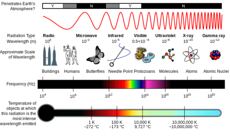
Light is one form of electromagnetic radiation. As such, it occupies a small slice of the electromagnetic spectrum, which also includes forms of radiation having greater energy than visible light, such as gamma rays and x-rays, and other forms having less energy than light, such as infrared radiation and microwaves. Each of these is characterized by two physical parameters that describe the quality of radiation: wavelength (λ) and frequency (ν). Visible light includes radiation having a wavelength between approximately 400 nm and 700 nm. Because the speed of radiation, c, is a universal constant, wavelength and frequency always vary inversely with each other and their product is equal to c.
A diagram of the Milton spectrum, showing the type, wavelength (with examples), frequency, the black body emission temperature.
Because light is a form of energy, and because we are interested in understanding how light energy is transduced into chemical energy, it is important to understand the relation between light quality and energy. Comparing blue light (450nm < λ < 475nm) with red light (620nm < λ < 730nm) for example, note that blue light has a smaller wavelength and therefore a greater frequency than red light. Even though the speed of light is a constant, the energy is not — energy varies in direct relation to frequency. So in the above example, blue light contains greater energy than red light of a given fluence rate.
In addition to being described in terms of its wave-like properties, the energy in light exists in discrete packets (called photons) rather than as a continuous stream. Photons are measurable down to the individual one, known as a quantum. As we will see next, the fact that light travels in photons, each having a predictable quantum of energy, helps us understand the events surrounding light absorbance by molecules and the transduction of energy into a chemical form.
Light Interacts with Molecules
As you look around the world, you see objects of all different colors. The production of those colors, and in fact our ability to perceive them, both rest on the fact that visible light can interact with matter in specific, predictable ways. Whether a particular frequency of radiation interacts with a particular molecule depends on the structure of the molecule — the arrangement of atoms in space. In the same way an aerial antenna depends on the precise spacing of its elements to intercept radio waves (another form of electromagnetic radiation) of specific frequencies, so too are biological molecules ‘tuned’ to specific frequencies of light to maximize energy transduction. In the case of the photosynthetic pigments known as chlorophylls, their molecular structure allows them to absorb blue and red wavelengths, but not yellow, green, or orange. While most plants have pigments that absorb yellow and orange, green light goes unused by the plant, being either transmitted or reflected.
When a pigment molecule intercepts a photon for which it is receptive, the energy of the photon is conserved by a rearrangement of electrons in the molecule. This rearrangement results in the boosting of an electron from the ground state to an excited state. Sometimes, the excited state is dissipated through the re-emission of light energy, known as fluorescence. Other times, if there are like molecules in close proximity, the excited state can be passed among them through resonant energy transfer. Under certain highly specialized circumstances, the excited state can represent the first step toward a photochemical reaction in which the electron departs the excited state molecule and reduces a nearby electron acceptor. Finally, the excited state may be dissipated through a process called radiationless decay, which releases heat to the surroundings. Each one of these processes plays an important role in photosynthesis.
While not as persistent as gravity, plants regularly encounter touch stimuli in their environment. For twining plants, the presence of a solid support induces differential growth of the tendrils, resulting in the familiar coiling response and the association with the support. For roots, touch sensing takes the form of avoiding obstacles in the path of elongation, resulting in the redirection of the root around the object. For stems, touch sensing may mean the perception of wind and other forces that shake the stem, leading to its increased radial thickening and rigidity. Without this rigidification in response to mechanical stimuli, plants would probably collapse under their own weight.
Touch stimulation results in both rapid changes within cells and longer-lived changes in development. One of the first events observed after a touch stimulus is an increase in intracellular calcium ions (Ca2+). The release of Ca2+ is probably linked to the temporary mechanical deformation of the plasma membrane, which contains mechanosensitive ion channels. One popular model holds that these channels open upon a mechanical stimulus, thus allowing the influx of Ca2+ leading to the elevation of intracellular concentrations.
The elevated Ca2+ concentration within the cell induces two other rapid changes in response to touch. High Ca2+ inhibits the plasma membrane H+-ATPase and probably also stimulates the opening of inward rectifying H+ channels, both of which drive an increase in intracellular H+ levels and an increasing alkalinization of the apoplast. Elevated Ca2+ also stimulates the production of reactive oxygen species outside the cell by activating an enzyme in the plasma membrane. Both ROS and alkalinization of the apoplast are likely associated with increasing the rigidity of the cell wall as a result of mechanical stimulation. Interestingly, both the acidification of the cytoplasm and ROS production can be induced by artificially increasing Ca2+ levels.
Longer-term responses to touch involve the synthesis of the gaseous plant hormone ethylene, as well as the upregulation of a variety of genes, including the TCH genes. Several of the TCH genes encode Ca2+ binding proteins such as calmodulins or proteins like calmodulins, which bind Ca2+ and mediate downstream responses. Another of the TCH genes (TCH4) encodes a xyloglucan endotransglycosylase, an enzyme involved in modifying cell wall polysaccharides.
References
Monshausen GB, Bibikova TN, Weisenseel MH, Gilroy S (2009). Ca2+ regulates reactive oxygen species production and pH during mechanosensing in Arabidopsis roots. The Plant Cell 21: 2341–2356
Plants respond to unilateral light by growing toward the light source (in the case of shoots) or away from the light source (in the case of roots). This response, known as phototropism, has an action spectrum with a broad peak in the blue region, and a maximum at approximately 450 nm. A biochemical approach isolated several membrane-bound proteins whose phosphorylation was required for the phototropic response, implicating a common signal transduction component in the plant’s response to unilateral light.
Using a mutant screen in the model plant Arabidopsis, several novel mutants were isolated that showed non-phototropic hypocotyls (the nph mutants). Cloning and characterization of the nph1 mutant revealed it to encode a polypeptide with several intriguing domains. On one end of the protein was a kinase domain, responsible for phosphorylating a target substrate, and on the other end of the protein was a LOV domain. LOV, (for light, oxygen, and voltage sensing), is an evolutionarily conserved domain found in a number of organisms and involved in sensing one of these physical cues. The protein encoded by NPH1, which has been named PHOTOTROPIN1 (phot1), also binds a flavin mononucleotide that acts as a chromophore.
PHOT1 protein has been localized to the plasma membrane in cells of dark-adapted hypocotyl tissue. Upon exposure to blue light, PHOT1 undergoes autophosphorylation and is relocalized to the cytoplasm. One role of PHOT1 appears to involve interacting with the auxin efflux carrier ABCB19 and causing its inhibition through phosphorylation. When ABCB19 is inhibited, auxin accumulates in the tip of the stem where it undergoes lateral transport to the shaded side of the stem through an efflux carrier.
After germination, plant growth is characterized by a the urgent need for the seedling to reach the light. During this period of growth, the seedling is throwing all of its resources into finding light. This kind of development, known as skotomorphogenesis, is characterized foremost by rapid elongation of the shoot, which is coupled with minimal elongation or further development of the root system. The shoot elongation that occurs during this period is strongly gravitropic, as this is one of the few cues available to the young seedling for orienting its growth. In dicot species, the energy-rich cotyledons are protected throughout this process of rapid stem elongation by the formation of an apical hook that tucks the cotyledons downward. Finally, dark-driven development is characterized by the lack of pigment biosynthesis, due in large part to the requirement for light as a catalyst in the production of chlorophyll. The seedling that develops in the dark is said to be etiolated.
Upon exposure to light for the first time, a number of changes occur to the seedling. The most obvious change is in the rate of stem elongation, which immediately slows and gives way to radial expansion. This is a very rapid response, occuring within seconds of the seedling sensing blue light. Almost concurrently with this, the primary root begins to elongate and will go on to begin producing lateral roots over the ensuing days. No longer needed for protection, the apical hook straightens and the cotyledons begin expanding and becoming green due to the synthesis of chlorophyll and other pigments required for photosynthesis.
The activation of such dramatic change in the transition from dark- to light-grown development requires many factors including several photoreceptors and many downstream signaling components. The rapid inhibition of stem elongation is mediated by a blue-light photoreceptor called cry1 (for cryptochrome). Many of the other responses associated with light are mediated by the phytochromes.
To identify other signal components of the photomorphogenesis pathway, mutant screens have been employed. One such screen was carried out to identify mutants that developed as though they had been exposed to light even though they hadn’t. These mutants were designated the constitutive photomorphogenesis (cop) mutants. The cop1 mutant was shown to have a lesion in a gene encoding an E3 ubiquitin ligase that targets transcription factors for degradation. These transcription factors regulate light-dependent genes, and by targeting them for degradation, COP1 acts as a negative regulator of photomorphogenesis. Many of the other COP genes encode components of a signalosome involved in regulating the localization of COP1 to either the nucleus or cytoplasm.
Following gravity perception, the sedimentation of amyloplasts must be converted into cellular information. Foremost among the candidates thought to specify this information is the formation of an auxin gradient.
Several different experimental approaches have shown that an auxin gradient does form during gravitropism. Using a radiolabelled-IAA approach, many researchers have confirmed that an auxin gradient forms throughout the elongation zone of gravity-stimulated roots. One of the limitations of this approach is the need to isolate a significant quantity of tissue, making it difficult to correlate the formation of the auxin gradient within the cells thought to be sensing gravity at the cap. More recently, an auxin gradient has been detected across the root cap columella cells by using an auxin-responsive promoter fused to GFP. This approach allows for the visualization of the effects of auxin within the cell, namely the resulting expression of a gene having a synthetic auxin response element. Because the GFP reporter protein must be produced and folded, there is a lag of approximately 1.5 h before a gradient of GFP appears following gravistimulation.
Another line of evidence that links gravity sensing with auxin redistribution is the very rapid relocalization that occurs to PIN3 proteins following gravistimulation. In vertical roots, PIN3 is localized uniformly throughout the plasma membrane. Within minutes after reorientation, PIN3 becomes localized to the new lower face of the cell, where it presumably directs auxin flux toward the lower flank of the root and inhibits their elongation. This relocalization is not due to the production of new PIN3 proteins, but rather results from the re-uptake of PIN3 from the plasma membrane via endocytosis. This is followed by the targeted exocytosis of vesicles containing PIN3 to a specified face of the plasma membrane.
While both of these lines of evidence support a link between gravity perception and auxin transport, the connection between the sedimentation of amyloplasts and redirection of PIN proteins remains loose. By crossing the auxin-responsive GFP into starchless mutants, we have shown that starchless roots fail to produce an auxin gradient across the root cap. This further supports the link between statolith sedimentation and the control of auxin transport.
In addition to auxin, several other potential growth regulating signals have been implicated in gravity signaling. For example, one of the fastest changes to be observed in gravity sensing cells is a change in ionic currents, measured as both membrane potential changes and surface potential changes. These changes have been reported within seconds after gravistimulation. Similarly, changes in pH have also been measured within minutes of gravistimulation, with root cap columella cells becoming alkaline and the apoplasts of these cells becoming acidic as a result of proton pumping. As of now, it is unclear whether or how these signals carry information about polarity that influences growth.
When a plant organ detects, through starch statolith sedimentation or another means, that it is no longer oriented in its preferred direction relative to gravity, a series of cellular and molecular events is initiated that results in a change in its direction of growth. This response, known as differential growth, leads to the eventual curvature of the organ back to its preferred orientation.
Some of the earliest studies of differential growth regulation focused not on gravitropic stimulation, but rather on phototropic stimulation. Charles Darwin was among the first plant scientists to study this phenomenon carefully:
…when seedlings are freely exposed to unilateral light, some influence is transmitted from the upper to the lower part, causing the latter to bend…
Charles Darwin, The Power of Movement in Plants, 1881
This summary came after careful experiments in which he shaded (or painted!) the tips of the growing seedlings and concluded that, while light seemed to be mostly perceived near the growing tip, the response occurred farther down the stem. Thus was set off a search for the nature of the “influence” that was transmitted, eventually leading to the discovery of the plant hormone auxin.
In a series of follow-up experiments to Darwin’s, it was shown that the factor causing differential growth could be extracted from rapidly growing tissues by placing tissue segments on a block of gelatin. The gelatin could then be used as a source of growth factor, and differential growth could be induced by applying the gelatin to one side of intact seedlings. These experiments suggested that differential growth begins with an unequal distribution of auxin, a concept known as the Cholodny-Went theory, first proposed in 1927.
A significant collection of physiological and biochemical evidence accumulated in support of the Cholodny-Went theory, including the discovery that auxin is transported in a polar manner through many organs of the plant. These data pointed to the need for a cell to have a mechanism for controlling the direction of auxin transport. This mechanism has now been identified to consist of several different kinds of auxin transporters, including the efflux carriers (PINs and ABCBs) and influx carriers (AUX1 and LAX3).
Immediately following germination, as the root emerges from the seed, it is faced with a decision: which way to grow? From the standpoint of adaptation, roots need to find a source of water and minerals quickly in order to sustain growth, both their own and that of the emerging shoot. Because water always flows downhill, the question of which way to grow seems to be reducible to, “Which way is down?”
In order for the plant to detect its position in a gravity field, it must have some way to transduce the acceleration force due to gravity into cellular information. Most evidence points to the change in position of starch-filled plastids (called amyloplasts) within the cell as playing a central role in sensing the position of the organ in the gravity field. As an organ elongates and becomes displaced from vertical, these amyloplasts sediment to the new lower side of the cell due to their high density relative to the cytoplasm. These amyloplasts, therefore, act as statoliths, and their role in sensing gravity is known as the starch-statolith hypothesis.
Root tips of wild type and starchless mutants showing the presence or absence of starch statoliths in the columella cells of the cap.
There are several strong pieces of evidence to support the starch-statolith hypothesis. Plants that are unable to synthesize starch due to a mutation in a gene encoding a key enzyme in that process show a significant reduction in gravity responses, even though their growth rates are nearly wild-type. Similarly, mutants with intermediate levels of starch biosynthesis show gravity responses that correlate with the amount of statolith movement. Thus, full gravity sensitivity seems to require a full complement of starch within the amyloplasts to provide the density necessary for statolith sedimentation.
If the movement of statoliths within the gravity-sensing cells is sufficient to induce a growth response, then causing the movement of the statoliths by some other means besides gravity stimulation should induce a growth response as well. In a clever series of experiments, one team of researchers accomplished just such displacement of statoliths in vertically-growing roots. By taking advantage of the diamagnetic properties of starch, coupled with the ability to displace a diamagnetic material in a high-gradient magnetic field (HGMF), they induced root curvature similar to gravitropic growth by ‘pushing’ the statoliths within the cell.
A final kind of evidence implicating statolith sedimentation in gravity sensing comes from experiments in which the tissue containing the statoliths is somehow disturbed. The earliest reports of this kind of disturbance date to the mid-19th century. In roots of some species, the root cap is relatively easy to surgically remove without damaging the meristem. Upon removal, the root loses its ability to respond to gravity, and it slowly regains it as the cap is replaced through continued cell divisions. In a much more modern interpretation of this decapping experiment, investigators used laser ablation to obliterate specific tiers of the columella cells in the root cap, concluding that much of the gravity sensing appears to reside in cells in the second and third tiers of the columella.
Unlike in the root, where much gravity sensing appears to be localized to the root cap columella cells, sensing is distributed throughout the growing region of the young shoot. In dicots, sedimenting amyloplasts are found in the endodermis, a cyclinder of tissue throughout the stem just outside the vascular cylinder. In monocots, which produce a protective sheath called a coleoptile that encloses the emerging leaf, amyloplasts are distributed throughout the sheath.


 Immediately following germination, as the root emerges from the seed, it is faced with a decision: which way to grow? From the standpoint of adaptation, roots need to find a source of water and minerals quickly in order to sustain growth, both their own and that of the emerging shoot. Because water always flows downhill, the question of which way to grow seems to be reducible to, “Which way is down?”
